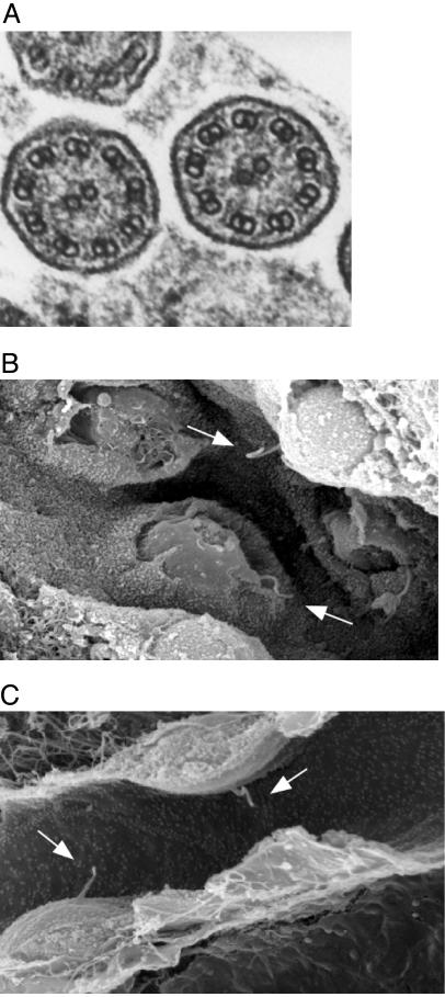
Fig. 5.
Electron microscopy analysis of motile and primary cilia. (A) Transmission electron microscopic image of a cross-section of cilia in the trachea of a Bbs4–/– mouse. The nine outer and two inner microtubules are clearly defined, indicating that the internal structure of motile cilia appears normal. (B and C) Scanning electron microscopic image of renal tubule cells from a Bbs4+/+ (B) and a Bbs4–/– (C) mouse. Normal appearing primary cilia (arrows) are evident in both.

