Fig. 1.
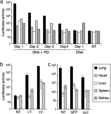
PEI promotes DNA and siRNA uptake in the lungs. (a) PEI mediates DNA transfection in the lungs after i.v. injection. Luc-expressing DNA (60 μg) was mixed with PEI at an N/P ratio of 10. The mixture was then injected into mice i.v. As controls, mice were not injected with anything (NT, no treatment) or injected with the same amount of naked DNA. One, 2, 3, and 4 days after injection, the indicated organs were harvested and homogenized. Luc activity and amount of protein in the homogenates were assayed. Shown are Luc activities in relative light units in 0.5 mg of protein in different organs over 4 days. Data shown are from one of two experiments. (b) PEI mediates DNA transfection in the lungs after i.t administration. The assays were done in the same way as in a, except that some mice were given DNA–PEI complexes i.t. Shown are average Luc activities in 0.5 mg of protein in the indicated organs of three mice per group 24 hr after DNA administration. Error bars indicate standard deviation. (c) PEI promotes siRNA uptake in the lungs after i.v. administration. Mice were given Luc-expressing DNA (60 μg)-PEI complexes i.t. and then promptly injected i.v. with siRNA–PEI complexes. Sixty micrograms of GFP- or Luc-specific siRNAs was used per mouse (at an N/P ratio of 5). For the NT group, mice were given the same volume of 5% glucose. Twenty-four hours later, Luc activity was assayed in homogenates of lungs, liver, and spleen. Shown are average Luc activity values in 0.5 mg of protein for three mice per group. Error bars indicate standard deviation.

