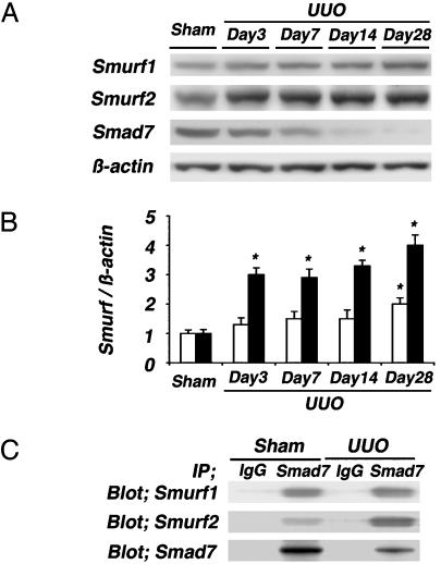
Fig. 5.
Immunoblot analyses of Smurf1 and Smurf2. (A) Western blot analyses of Smurf1 and Smurf2 were performed in sham-operated and UUO kidneys on days 3, 7, 14, and 28. (B) Densitometric analysis of the levels of Smurf1 and Smurf2 detected by Western blot analysis. In UUO kidneys, marked increases in Smurf2 (filled bars) were noted at day 3 and further enhanced thereafter in almost-inverse proportion to the levels of Smad7, whereas the increases in Smurf1 (open bars) were milder than the increases of Smurf2. (C) Renal extracts collected from sham-operated and UUO kidneys on day 7 were subjected to Smad7 immunoprecipitation (IP), followed by Smurf1 and Smurf2 immunoblotting (Blot). In UUO kidneys, both the Smad7–Smurf1 and Smad7–Smurf2 complexes were noted. The levels of Smurf2–Smad7 complex were relatively more than those of Smurf1–Smad7 complex. Representative data of three independent experiments are shown. Data are given as mean ± SEM values of nine mice in each group. *, P < 0.05, compared with sham-operated kidneys.

