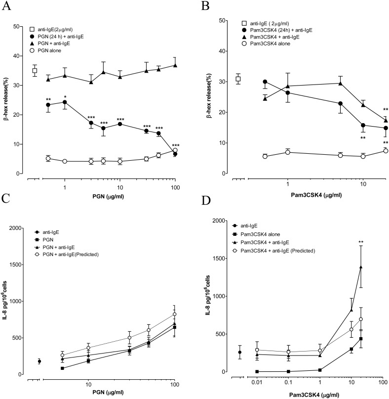
Figure 1. Effect of TLR2 ligands on anti-IgE induced degranulation and IL-8 release from LAD2 cells.
(A, B) LAD2 cells were incubated with only PGN or Pam3CSK4 for 30 min (○). LAD2 cells were incubated with anti-IgE (2 µg/ml) at the same time (▴) or after 24 h pre-incubation (•) with PGN or Pam3CSK4. The levels of β-hex release induced by anti-IgE alone and in the present of TLR2 ligands were compared with one-way ANOVA and Dunnett’s multiple comparison tests. *p<0.05, **p<0.01, ***p<0.001 (n = 3–5). (C, D) LAD2 cells were incubated alone with anti-IgE (2 µg/ml, •), PGN/Pam3CSK4 (▪) or combination of anti-IgE with PGN/Pam3CSK4 (▴) for 24 h. Two-way ANOVA and Bonferroni posttests were applied to compare the actual amount of IL-8 released with the predicted value obtained by adding the individual amounts released by anti-IgE and PGN or Pam3CSK4 (○). **p<0.01 (n = 5).

