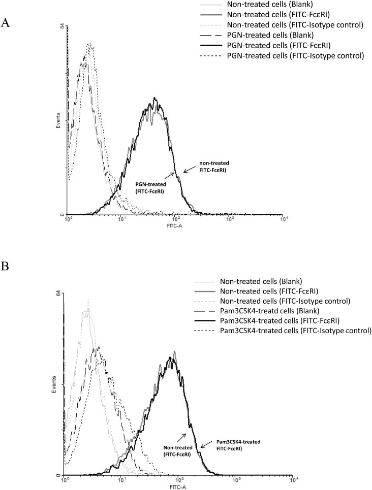
Figure 2. Effects of PGN and Pam3CSK4 on the expression of FcεRI on LAD2 cells.
LAD2 cells (without IgE sensitization) were incubated with PGN (50 µg/ml) (A) and Pam3CSK4 (20 µg/ml) (B) for 24 h and FcεRI surface expression was analyzed by flow cytometry after cells were incubated with FITC-conjugated anti-human FcεRI antibody, FITC-conjugated mouse IgG2b isotype control or FACS buffer for specific labelling of FcεRI, isotype and blank control respectively. The FcεRI expression of cells that were not treated (grey curve) or treated (blank curve) with the TLR2 ligands was not different as shown. Results were representative of four independent experiments.

