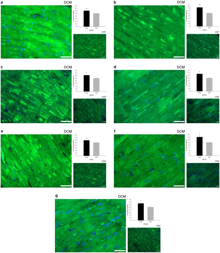
Figure 2. Mitochondrial protein overexpression in dilated human hearts according to immunofluorescence techniques.
Influence of dilated cardiomyopathy on the amount of each representative protein involved in cardiac energy metabolism (ODPA, ETFD, DLDH, AL4A1, and ATPA), protein biosynthesis (EFTU), and the stress response (PRDX3). Immunofluorescence of (a) ODPA, (b) ETFD, (c) DLDH, (d) AL4A1, (e) ATPA, (f) EFTU, and (g) PRDX3 were significantly increased in patients with dilated cardiomyopathy compared with the control group. Here we show the nucleus co-stained with DAPI (blue). All of the micrographs are representative of the results obtained in four independent experiments for each group and protein studied, DCM (n = 4) and CNT (n = 4). The bar represents 100 µm. The bar graph shows the relative fluorescence intensity in dilated compared to control hearts. The data are expressed as mean ± SEM. CNT, control; DCM, dilated cardiomyopathy; ODPA, pyruvate dehydrogenase E1 component subunit α, somatic form; ETFD, electron transfer flavoprotein-ubiquinone oxidoreductase; DLDH, dihydrolipoyl dehydrogenase; AL4A1, delta-1-pyrroline-5-carboxylate dehydrogenase; ATPA, ATP synthase subunit α; EFTU, elongation factor Tu; PRDX3, thioredoxin-dependent peroxide reductase. *p value<0.05, **p value<0.01.

