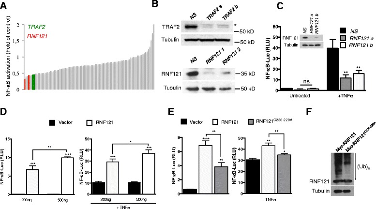
Figure 1.

Participation of RNF121 in TNFR-mediated NF-κB activation. (A) NF-κB reporter luciferase assay screen of a siRNA library targeting 46 transmembrane E3 ubiquitin ligases (2 siRNAs/target) in HEK293T cells. Cells were stimulated with TNFα (10 ng/ml) for 6 hrs and fold activation compared to non-specific (NS) siRNA-treated cells was calculated. Red and green histograms indicate siRNA against RNF121 and TRAF2, respectively. TRAF2 was used as a positive control. (B) Cell extracts from HEK293T cells transfected as in (A) were analyzed by immunoblot as indicated. (C) HEK293T cells transfected with a control non-specific (NS) siRNA or with siRNAs against RNF121 (RNF121 a or b), were also transfected 48 hrs later with an NF-κB reporter. 24 hrs later, the cells were either left unstimulated or were stimulated with TNFα (10 ng/ml) for 6 hrs and then were analyzed by luciferase assay. The results were normalized against Renilla luciferase activity [analysis of variance (ANOVA)]. ns: not significant. RLU, Relative Light Units. Inset: Immunoblotting analysis of the knockdown of RNF121 by the specific siRNAs. (D) NF-κB reporter luciferase assay in HEK293T cells transfected with increasing concentrations (200 or 500 ng) of a Myc-tagged plasmid coding for RNF121 and left unstimulated (left panel) or stimulated with TNFα (1 ng/ml) for 6 hrs (right panel) [analysis of variance (Student’s t-tests)]. (E) NF-κB reporter luciferase assay in HEK293T cells transfected with 200 ng of a Myc-tagged plasmid coding for RNF121 or for the mutant RNF121C226-229A and left unstimulated (left panel) or stimulated with TNFα (10 ng/ml) for 6 hrs (right panel) [analysis of variance (Student’s t-tests)]. (F) HEK293T cells were transfected with a Myc-tagged plasmid coding for RNF121 or for the mutant RNF121C226-229A. 24 hrs later, cell extracts were analyzed by immunoblotting. (Ub)n indicates poly-ubiquitylated species.
