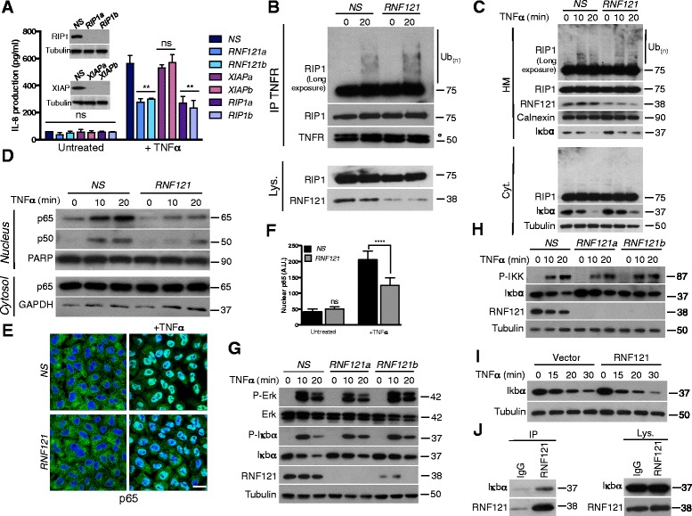
Figure 3.

RNF121 regulates NF-κB activation through IκBα degradation. (A) HEK293T cells were transfected with a control non-specific (NS) siRNA or with the indicated siRNAs. 72 hrs later, the cells were either left untreated or exposed to TNFα (1 ng/ml). The secretion of IL-8 was measured by ELISA. (B) HEK293T cells were transfected for 72 hrs with NS siRNA or with a siRNA against RNF121 (RNF121). Cells were then either left untreated or exposed to 10 ng/ml TNFα for 20 min. Cell lysates (Lys.) were subjected to immunoprecipitation (IP) with an antibody raised against TNFR. ° IgG heavy chains. (C) NS- and RNF121-silenced HEK293T cells were stimulated with TNFα (10 ng/ml) for the indicated times. Crude HM and cytosolic (Cyt.) fractions were analyzed by immunoblotting. (D) Nuclear and cytoplasmic extracts from cells stimulated as in (C) were analyzed by immunoblotting as indicated. (E) NS- and RNF121-silenced HeLa cells were either left untreated or exposed to TNFα for 20 min. Nuclear translocation of the p65 NF-κB subunit was assessed by immunofluorescence. Scale bar : 50 μm. The pixel intensity of the nuclear signal of p65 in each condition was quantified in (F). A.U.: Arbitrary unit. (G) NS- and RNF121-silenced HEK293T were stimulated with TNFα for the indicated times, then were subjected to immunoblotting analysis as indicated. (H) Same conditions as (G) but cells were pre-treated 5 min with the broad phosphatase inhibitor Calyculin A (50 nM) before TNFα stimulation. (I) HEK293T cells were transfected with a control empty vector or Myc-tagged plasmid coding for RNF121. 24 hrs later, cells were stimulated with TNFα for the indicated times and cell extracts were analyzed by immunoblotting as indicated. (J) Cell lysates (Lys.) from HEK293T were subjected to immunoprecipitation (IP) with an antibody raised against RNF121 or with a control IgG.
