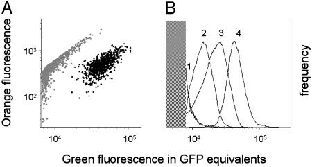
Fig. 2.
In vivo GFP expression from single-copy transcriptional fusions to chromosomal Salmonella promoters. (A) Two-color flow cytometry of spleen homogenate of a mouse infected with SL1344 sifB::gfp. Gray dots represent autofluorescent host tissue fragments, and black dots represent GFP-containing Salmonella cells (17). (B) Comparison of GFP expression from various chromosomal gfp fusions in infected spleen (1, SL1344 yjiS::gfp; 2, SL1344 mig14::gfp; 3, SL1344 nt01st5349::gfp; and 4, SL1344 virK::gfp). The shaded area represents background autofluorescence preventing GFP detection. Only a small bright fraction of Salmonella strain 1 was detectable.

