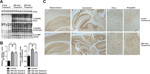
Figure 2.

O -GlcNAc levels are increased in the TAPP mouse brain. A. Western blots of total brain homogenates from 0, 200 and 500 mkd Thiamet-G treated TAPP mice reveals that O-GlcNAc levels are vastly increased (RL2 and CTD110.6) while actin indicates equal protein loading. B. Quantification of O-GlcNAc immunoreactivity (CTD110.6) normalized to actin by densitometry of all bands (left panel) or only the low molecular weight (MW) bands (bands <50 kDa, right panel). N =10 in each group. *indicates p <0.05, ***indicates p <0.001, unpaired two-tailed t-test) C. Immunohistochemical (IHC) analysis of 0 and 500 mkd Thiamet-G treated TAPP mice brain tissue reveals that O-GlcNAc levels are increased in all of the hippocampus (A’, E’), cerebellum (B’, F’), pons (C’, G’) and the amygdala (D’ , H’).
