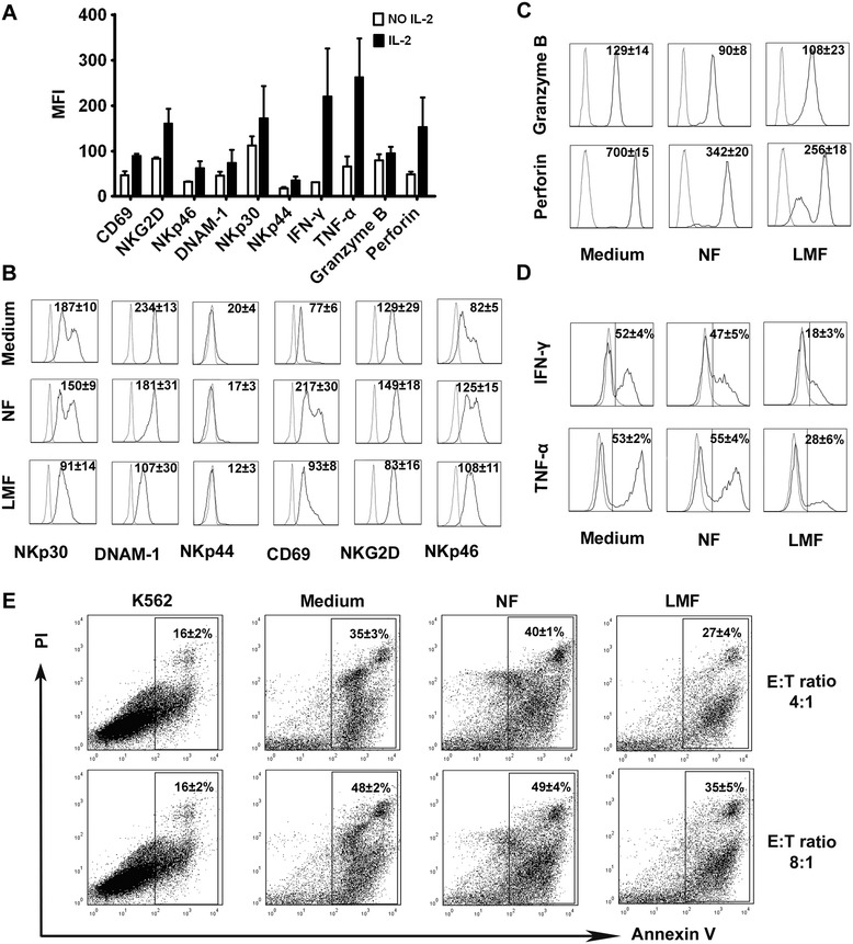
Figure 3.

LMFs regulate NK cell function. NK cells were cultured in rhIL-2 alone or with the indicated fibroblast cells and analyzed by flow cytometry. (A) After 5 days of culture in the absence or presence of rhIL-2, the triggering receptors, cytolytic granules and cytokine production of NK cells were analyzed. (B) The expression of NK cell triggering receptors was analyzed. (C) Intracytoplasmic analysis of granzyme B and perforin expression. (D) Analysis of the production of IFN-γ and TNF-α by NK cells under different conditions. (E) LMF-conditioned NK cells showed reduced cytotoxicity against K562 cells at different T/E ratios. The mean fluorescence intensities (MFI; indicated as the mean ± SEM of 7 independent experiments; A, B and C) and the percentages of positive cells (D) or apoptotic cells (E) are shown. The open profiles with dotted lines show the isotype control, and the open profiles with solid lines show the expression of the indicated markers.
