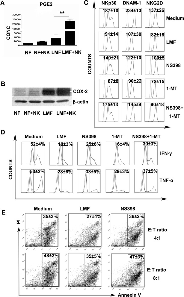
Figure 4.

Role of PGE2 in the immunoregulatory effect of LMFs on NK cells. NK cells were co-cultured with LMFs in the absence or presence of the indicated inhibitors for 5 days with rhIL-2. (A) NK cells were left untreated except for rhIL-2 (NK) or were cultured with the indicated fibroblasts (NF, normal skin fibroblasts; LMF, LMFs) for 5 days. The concentrations of PGE2 (pg/mL) in the supernatants were assessed by ELISA. (B) The expression of COX-2 by the above-mentioned cells were assessed by Western blotting. (C) The variations in NKp30, DNAM-1 and NKG2D in the presence of the indicated inhibitors. (D) The secretion of IFN-γ and TNF-α by NK cells was affected by inhibitors. (E) LMF-conditioned NK cells showed restored cytotoxicity against K562 cells with the inhibitors. The mean fluorescence intensities (MFI; indicated as the mean ± SEM of 7 independent experiments) (C) and the percentages of receptor-positive cells (D) or apoptotic cells (E) are shown. The open profiles with dotted lines show the isotype control, and the open profiles with solid lines show the expression of the indicated markers. *P<0.05, **P<0.01.
