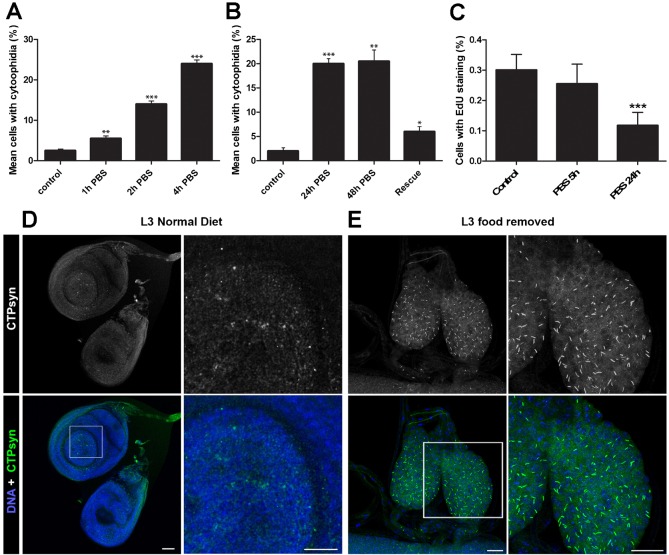
Fig. 1. Cytoophidium assembly occurs in response to nutrient stress.
(A) Mean number of cells containing visible cytoophidia increases in nutrient restricted cells. Significantly more cytoophidia are seen in S2R+ cells in PBS after just one hour. Control cells are maintained in complete Drosophila Schneider's medium. At least 100 cells were counted from ten sites per well from at least three independent replicates. (B) Cells with increased numbers of cytoophidia following nutrient restriction can be rescued by addition of whole media. Cells were examined two hours after re-introduction of whole media. (C) No significant change in EdU marked cells is observed after 5 hours, indicating that cell cycle arrest is not necessary for cytoophidia formation in S2 cells. (D) Response to nutrient restriction in imaginal disc tissue in vivo. Imaginal discs of L3 larvae display only diffuse cytoophidia when fed a normal diet. (E) In larvae raised on a nutrient restricted diet, the imaginal discs are smaller and cytoophidia are highly prevalent. Scale bars: 10 µm. ***P<0.001, **P<0.01, *P<0.05.

