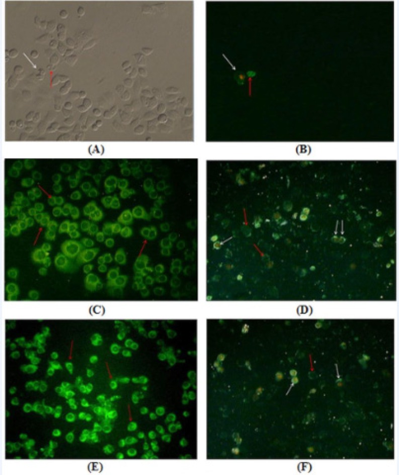
Figure 3.
Annexin V–FLUOS and PI staining of AGS cells treated with F. angulata Boiss. leaf and flower extracts for 24 h. Early apoptotic cells with only Annexin V–FLUOS positive staining were recognized by green plasma membrane (red arrows), while late stage apoptotic cells with both Annexin V–FLUOS and PI positive staining were observed with green membrane and red nucleus (white arrows). A) and B) Untreated AGS cells observed under inverted microscope and fluorescence microscope, respectively. C) and D) AGS cells treated with 80 μg/mL and 160 μg/mL concentrations of F. angulata Boiss. leaf extracts, respectively. E) and F) AGS cells treated with 80 μg/mL and 160 μg/mL concentrations of F. angulata Boiss. flower extracts, respectively. Magnification 200X

