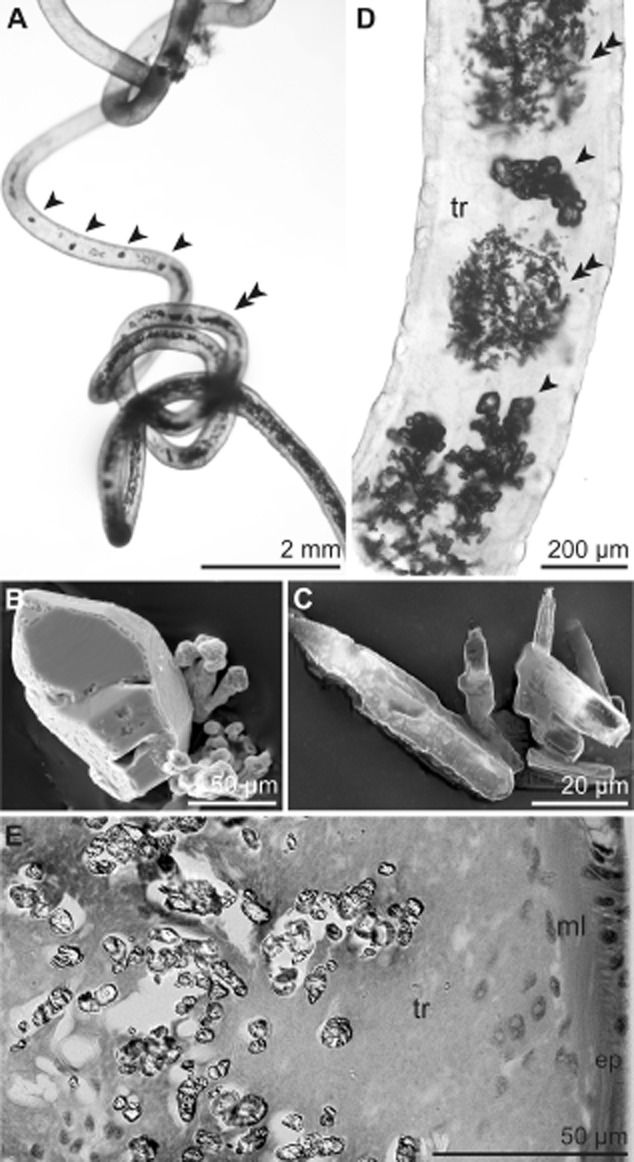
Fig 3.

Crystals deposited in the S. contortum trophosome. A. Whole specimen within the tube viewed under a dissecting microscope containing orthorhombic (arrowhead) and needle-shaped crystals (double arrowhead). B–C. SEM of orthorhombic (B) and needle-shaped crystals (C). D. LM of whole mount of the posterior body region showing regions of densely packed needle-shaped crystals (double arrowhead) interspersed by orthorhombic ones (arrowhead). E. LM of high-pressure frozen and freeze-substituted sample of the posterior trophosome. Crystals are limited to the trophosomal tissue located in the body cavity of the trunk. Ep, epidermis; ml, muscle layer; tr, trophosome.
