
Figure 1.
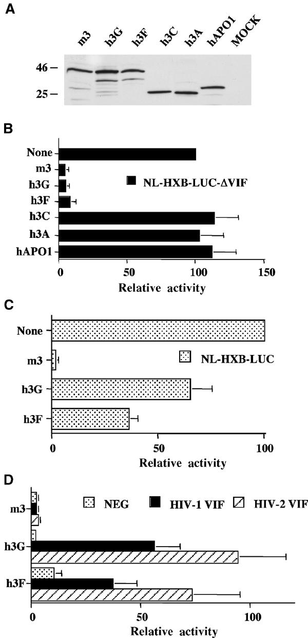
Inhibition of productive HIV-1 infection by h3F is suppressed by both HIV-1 and HIV-2 Vif. (A) Western analysis of HA epitope-tagged forms of the indicated APOBEC proteins expressed in transfected 293T cells. (B) 293T cells were transfected with pNL-HXB-LUCΔVIF (1.5 μg) and the indicated APOBEC expression plasmid (125 ng). At 44 h after transfection, supernatant media were collected and used to infect CD4+ CXCR4+ 293T cells. A further 28 h later, these cells were lysed and induced luciferase activities were quantified. The average of three independent experiments with standard deviation is indicated. Activities are given relative to the virus obtained from the culture transfected with pNL-HXB-LUCΔVIF and the parental pcDNA3 plasmid, which was set at 100. (C) Similar to panel B, except that the Vif+ pNL-HXB-LUC indicator virus was used. (D) Similar to panel B, except that the cells were additionally transfected with 250 ng of a plasmid expressing HIV-1 Vif or HIV-2 Vif, or the pgΔVif plasmid as a negative control.