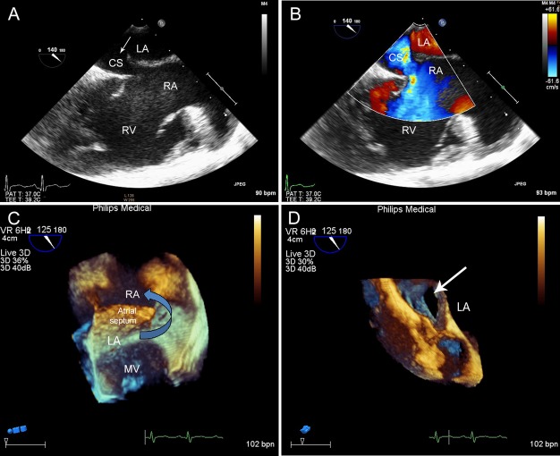
Figure 1.
Imaging results of patient no. 9 (an 18-year-old female with PUCS). En face view of the CS showed the roof of the CS, which beyond the realm of the atrial septum, was partial absent. A. A shunt from the LA to the dilated CS, and ultimately to the RA was confirmed by the color Doppler flow imaging. B. In the 3DTEE image, the arrow shows that the LA is communicating with the RA through the defect. C. The white arrow points to the en face view of the defect. D. PUCS, partially unroofed coronary sinus; LA, left atrium; CS, coronary sinus; RA, right atrium; RV, right ventricle; MV, mitral valve.

