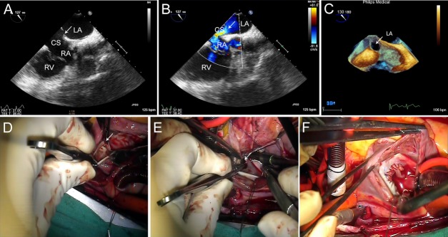
Figure 5.

Patient no. 6 (a 42-year-old-female with PUCS). En face view of the CS showed the roof defect. A, and a left-to-right shunt was confirmed by the color Doppler flow imaging. B. In the 3DTEE image, the white arrow points to PUCS. C. Surgical view from the RA; the white arrows point to the defect. D. Autologous pericardium was used to perform the roof repair. E–F. PUCS = partially unroofed coronary sinus; RA = right atrium.
