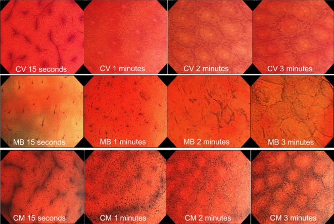
Figure 4.
Typical examples of endocytoscopic images stained with 0.05% crystal violet (CV) alone, 1% methylene blue (MB) alone and CV plus MB (CM double). With CV alone, nuclei were not recognized and gland formation reached ‘recognizable’ in 1 min. With MB alone, both nuclei and gland formation reached ‘recognizable’ in 2 min. With CM double, both nuclei and gland formation reached ‘recognizable’ in 1 min.

