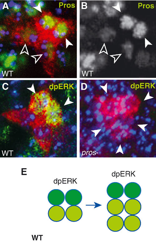
Figure 5.

No activation of ERK in LG in pros mutants. LG-lacZ visualised with anti-βgal (red). (A, B) Wild type: (A) high levels of anti-Pros (green, white arrowheads) in two cells and lower levels in the posterior two (empty arrowheads); (B) single-channel view showing only anti-Pros. (C, D) anti-dpERK (green, arrowheads) in the two LG with higher Pros levels: (C) in wild type; (D) no dpERK (arrowheads indicate larger cluster of LG at stage 12) is detected in pros mutants. (E) Diagram illustrating the segregation of dpERK (dark green) among the Pros-positive LG (green). All images represent one hemisegment, blue is TOTO-3, midline to the left, anterior up.
