
Figure 4.
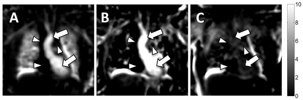
Coronal reformats of 3D OE maps generated from (A) TE = 0.08 ms and (B) TE = 2.1 ms acquisitions. Image quality and percent change in the lung parenchyma are poor at the longer echo time in this 3D radial acquisition due to the very short T2* environment. Enhancement of the oxygenated blood in the left ventricle and ascending aorta remains visible at both echo times (arrows). Note the lack of signal in the deoxygenated superior vena cava, right atrium, and pulmonary artery (arrowheads). (C) As a negative control, an OE map between two acquisitions obtained while breathing room air shows no oxygen enhancement in lung parenchyma or blood pool.