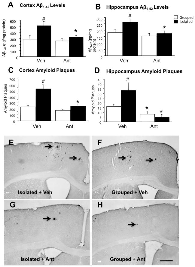
Figure 2. Antalarmin reduces tissue Aβ1-42 levels and Aβ plaque deposition in chronically stressed Tg2576 mice.
Tissue Aβ levels. Chronic isolation stress significantly increased Aβ1-42 levels as compared to grouphoused animals in the cortex and hippocampus (A, B: #P< .05) at ten months of age. However, chronic administration of antalarmin (20 mg/kg) significantly decreased Aβ1-42 levels as compared to the vehicle in isolated mice (A, B: *P< .05).
Aβ plaque deposition. The number of Aβ immunohistochemical positive plaques in the cortex and hippocampus (C, D: ## P< .01, #P< .05) were significantly increased in isolated mice as compared to group-housed mice. Antalarmin administration (20 mg/kg) significantly decreased Aβ plaque deposition in the cortex of isolated mice (C: *P<.01: Isolated +Ant versus Isolated +Veh) and decreased Aβ plaque deposition in the hippocampus of both isolated mice; and group-housed mice (D: *P< .05, Isolated +Ant versus Isolated +Veh; ★P<.05: Grouped +Ant versus Grouped + Veh).
Representative Aβ-immunocytochemical stained sections are shown from the cortex of 10-month-old Tg2576 mice from housed in isolation (Isolated, E, G) or under group-housed conditions (Grouped, F, H) and treated with antalarmin. Arrowheads indicate the plaques. Scale Bar =200 μm shown in panel H also applies to panels E, F, and G.

