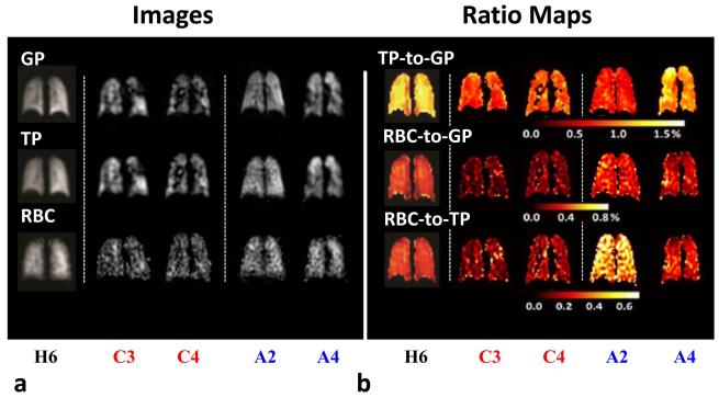
Figure 6.
(a) Images of HXe lung ventilation and HXe dissolved in TP and RBC compartments. (b) Corresponding TP-to-GP, RBC-to-GP and RBC-to-TP ratio maps acquired in healthy subject H6, COPD subjects C3 and C4, and asthma subjects A2 and A4. COPD subjects C3 and C4 showed numerous ventilation defects and had all ratios lower than those for the healthy subjects. Younger asthma subject A2 showed high RBC-to-TP ratios, as compared with healthy subjects. Similar to COPD subjects C3 and C4, the older asthma subject A4 had all ratios lower than those for the healthy subjects. In the apex of the right lung, this subject had high TP-to-GP ratios and low RBC-to-GP and RBC-to-TP ratios. Images adapted from (29) with permission.

