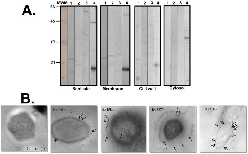
Figure 2.
Lipoprotein recognition. (A) Western blot with rabbit pre-immune serum and post-third inoculation against M. tuberculosis H37Rv total sonicate and subcellular fractions (membrane, cell wall and cytosol). Lanes 1 to 4 show post-third immune serum. Serums were obtained by inoculating polymeric peptides, as mentioned in the Materials and Methods section: (1) Rv1411c; (2) Rv1911c; (3) Rv2270 and (4) Rv3763. The molecular weight marker is shown on the left-hand side of the Figure. (B) Immunoelectron microscopy. 6,000 x magnification. Lipoprotein detection can be seen on M. tuberculosis H37Rv bacillus surface, as indicated by the colloidal gold particles indicated by black arrows.

