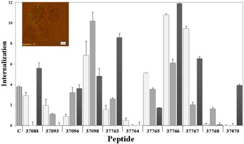
Figure 7.
Percentages of peptide–coated microspheres internalised by A549 cells. A549 cells were independently incubated with peptides and then with uncoated microspheres as control treatment (subtracted from the final result for each peptide). Untreated microspheres were used as negative control (C). The results were the average internalisation calculated for each treatment ± SD.* p ≤ 0.01; **p ≤ 0.001, according to a two-tailed Student's t-test. Insert shows the fluorescence microscopy of latex beads internalised by A549 cells.

