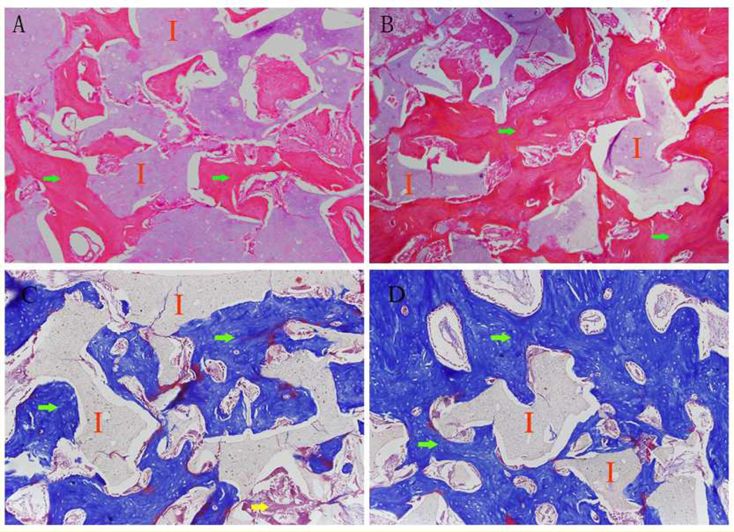
Figure 5.
Hemotoxylin and eosin (H&E) stained and Masson’s Trichrome stained sections of (A, C) Poly (octanediol citrate) – click – hydroxyapatite (POC-Click-HA) single-phase scaffolds and (B, D) POC-Click-HA biphasic-50 scaffolds after 15 weeks of implantation in a 10 mm long rabbit radial defect model showing the presence of new bone formation (I: implant; Green arrow: new bone formation; Yellow arrow: fibrous tissue; magnification 200×).

