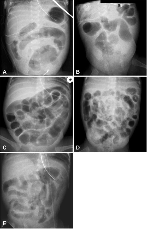
Figure 1.

Abdominal X-rays at clinical presentation of volvulus. Abdominal X-rays at clinical presentation of volvulus: Panel A shows possibly suggestive concentric alignement of bowel loops, Panels A, B, E display unspecific intestinal wall thickening, Panel D may well have been compatible with necrotising enterocolitis and Panel C is unspecific.
