Fig. 3.
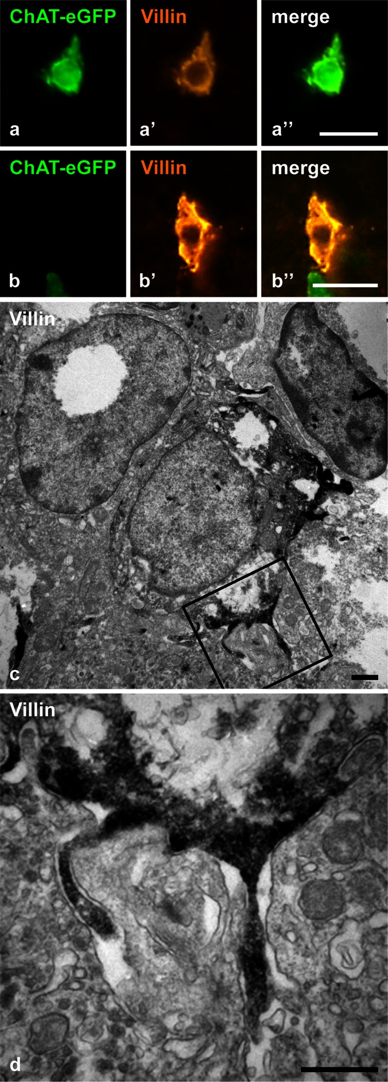
Villin immunolabeling. ChAT-eGFP-positive cell with immunoreactivity for villin (a–a”) and villin-positive cell lacking ChAT-eGFP-fluorescence (b–b”). c, d Ultrastructural immunohistochemistry, pre-embedding technique. Villin immunoreactivity is indicated by dark diaminobenzidine reaction product. A villin-immunoreactive epithelial cell extends lateral microvilli. d Higher magnification of boxed area in c. a Male aged 31 weeks. b Female aged 25 weeks. c, d Female aged 9 weeks. Bars 20 μm (a, b), 1 μm (c), 0.5 μm (d)

