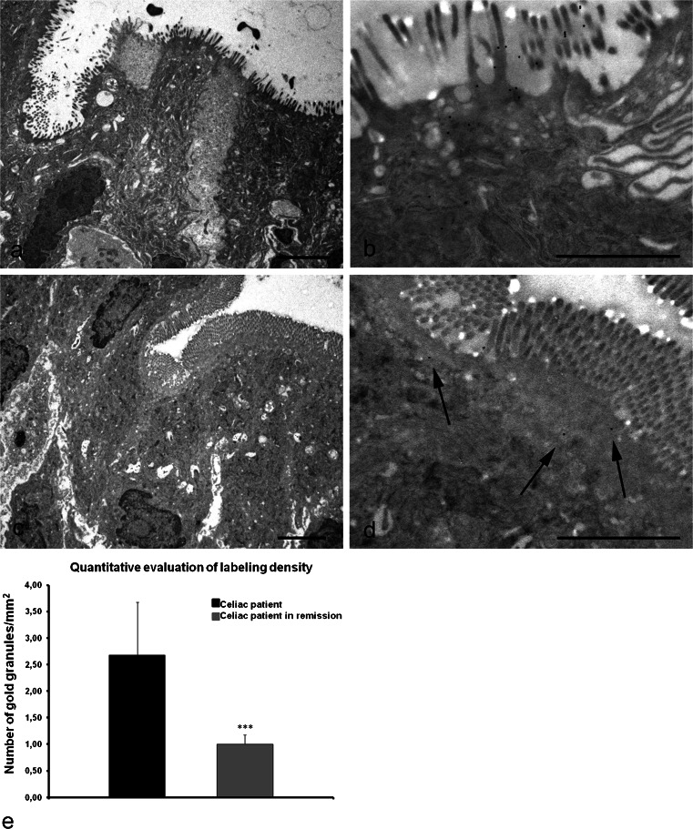
Fig. 7.
Electron micrographs of duodenal mucosa from celiac patients (a, b) and from celiac patients on a gluten-free diet (c, d). a Representative example of mucosal injury in a celiac patient: enterocytes display rarefaction and partial disappearance of microvilli. b Higher magnification of apical surface showing numerous gold granules: the presence of TG2 appears mostly to be associated with the microvilli on the luminal surface of enterocytes. c Ultrastructural features of enteric mucosa in a patient with celiac disease in remission after a gluten-free diet: a marked recovery of tissue architecture is visible, with virtually absent alterations in the brush border. d Higher magnification of the top surface facing the lumen: a change in the distribution of TG2 is demonstrated by the presence of just a few granules (arrows) localized underneath the microvilli. Bars 3 μm (a,c), 1.5 μm (b,d). e Quantification of TG2 immuno-gold labeling within the apical membranes of enterocytes in celiac patients and in celiac patients on a gluten free-diet. The bar chart shows the decrease in the number of gold particles per square micrometer within the apical surface of enterocytes of a patient in remission (gray) versus a diseased patient (black). Error bars are ± SEM. ***P < 0.0001

