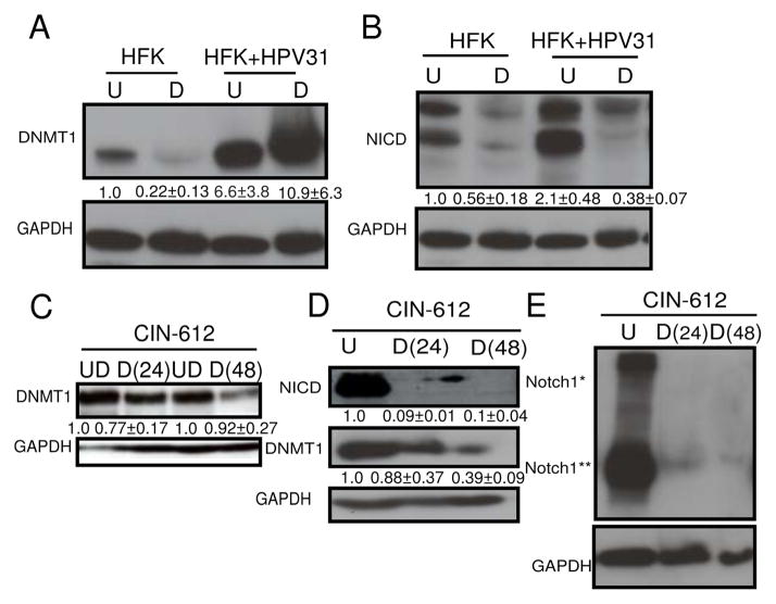
Figure 3. Human Papillomavirus 31 genomes enhance both Notch1 and DNMT1 levels.
A, Expression of DNMT1 in primary keratinocytes (HFK) compared to HFKs transfected with HPV31 genomes (HFK+ HPV31), in undifferentiated (U) and after differentiation (D) in methylcellulose. B, Expression of cleaved intracellular Notch1 (NICD) in HFKs compared with HFK+HPV31, in undifferentiated (U) and after differentiation (D) in methylcellulose. C, Expression of DNMT1 in undifferentiated CIN-612 cells (UD) and after differentiation in Ca2+ for 24 hrs [D(24)] or 48 hrs [D(48)]. D, Immunoblot of DNMT1 and NICD levels in CIN-612 cells in undifferentiated conditions (UD) and after differentiation in methylcellulose for 24 hrs [D(24)] or 48 hrs [D(48)]. E, Immunoblot of Notch1 levels in CIN-612 cells before (U) and after differentiation in methylcellulose for 24 hours [D(24)] and 48 hours [D(48)]. Notch1* represents full-length and Notch1** represents intracellular levels of Notch1. A–E, GAPDH was used as a loading control. N=3, mean with S.E.M. are shown below the blots.

