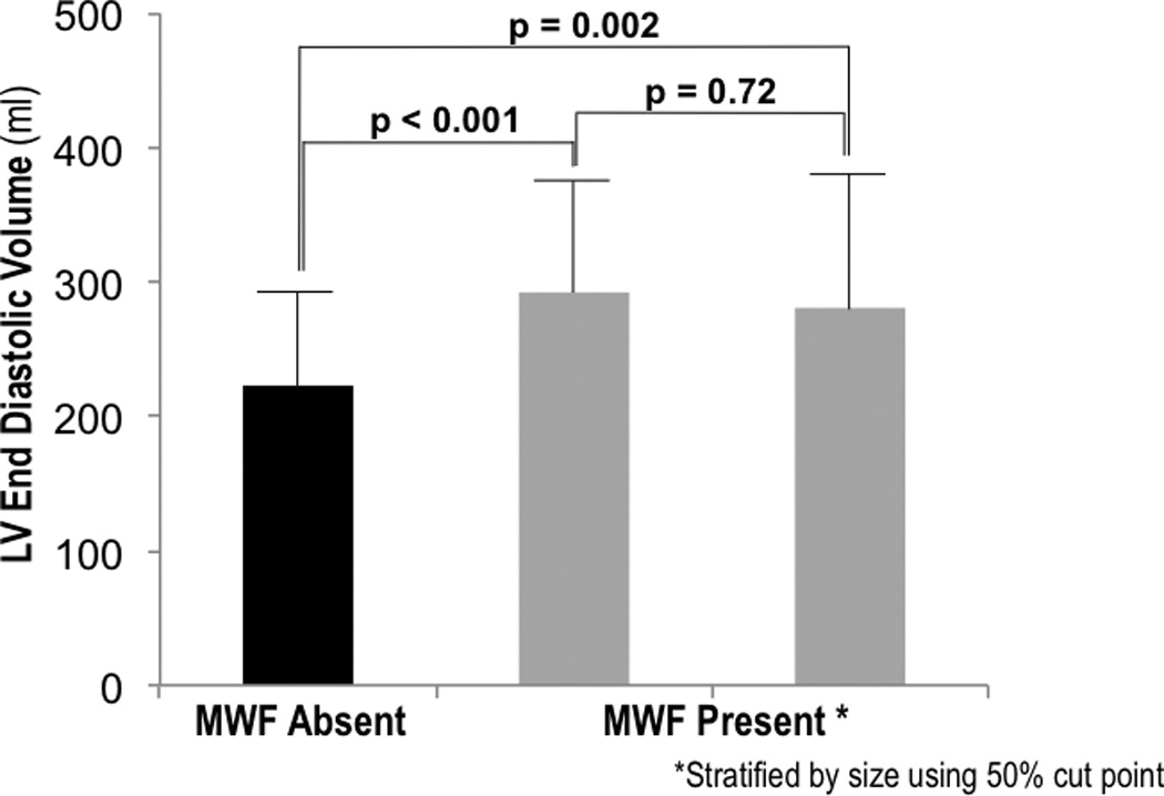
Figure 3. Mid-Wall Fibrosis Size in Relation to LV Remodeling.

LV end-diastolic volume (mean ± SD) among patients without MW-LGE (black bar), as well as MW-LGE-affected patients (grey bars) stratified into two groups based on fibrosis size (right = top, left = bottom 50%). Note that both groups of MW-LGE-affected patients had larger LV chamber volumes than did those without MW-LGE, with non-significant differences between MW-LGE-affected patients stratified by fibrosis size.
