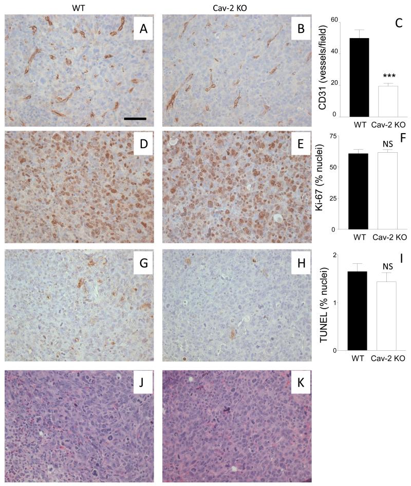
Figure 5. MVD versus cellular proliferation and death within the earliest palpable tumors extracted on day 6.
5-um paraffin sections from earliest palpable tumors extracted 6 days after s.c. implantation of LLC cells were immunohistochemically stained with antibodies against CD31 (A,B) and Ki67 (D,E), TUNEL kit (G,H) or histochemically stained with H&E (J,K) to assess MVD, proliferation, apoptotic/non-apoptotic death, and gross cellular morphology, respectively, C: Graphical representation of MVD within the tumors quantified based on CD31 positive staining with the data expressed as mean numbers of CD31 positive vessels per field ± S.E.M. ***, P<0.0001 compared with WT by unpaired t-test (n=7 for WT and n=14 for Cav-2 KO). F: Graphical representation of cell proliferation within tumors quantified based on Ki67 positive staining with the data expressed as mean numbers of Ki67 positive nuclei per field ± S.E.M. I: Graphical representation of cell death within tumors quantified based on TUNEL positive staining, with the data expressed as mean numbers of TUNEL positive nuclei per field ± S.E.M. NS, not statistically significant compared with WT by unpaired t-test (n=6 for WT for Cav-2 KO). Bar: 50 um.

