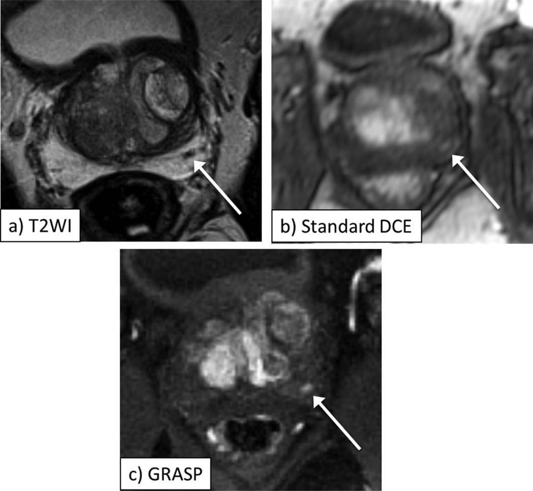
Figure 2.
A 63-year-old male with biopsy-proven Gleason 3+3 prostate cancer in the left midgland of the prostate, as depicted by area of decreased signal on axial T2-weighted image (a, arrow). Early postcontrast images from standard DCE (b) and GRASP DCE (c) show corresponding focus of abnormal early enhancement in this sextant (arrow, b,c), which is better defined on GRASP image. Also note more distinct visualization of anatomic details on GRASP image.

