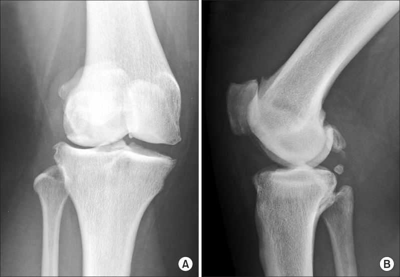
Fig. 1.
Preoperative radiographs of the right knee. (A) Anteroposterior view showing dense bone, narrowing of the lateral joint space, osteophytes in the lateral and medial plateau, and valgus alignment. (B) Lateral view showing dense bone, osteophytes at the patellofemoral joint and possible loose bodies.

