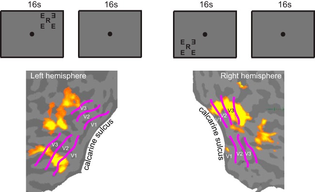
Fig. 2.

Defining the regions of interest (ROIs). An ROI was defined independently in a separate scan as the contiguous set of voxels within a retinotopically defined visual area (V1–V3) that were significantly activated by a counter-flickering stimulus. The stimulus consisted of a middle letter and 4 flankers simultaneously presented along the radial and tangential directions. The position of the middle letter, presentation timing, and the fixation task were identical to the main experiment. Two testing locations (upper and lower visual fields) were measured in the same scan, and the set of 2 stimulus conditions was repeated twice in a counterbalanced order. The activation maps show the V1, V2, and V3 ROIs determined for 1 subject. Retinotopic boundaries of V1, V2, and V3 were obtained with a standard retinotopic mapping method (Engel et al. 1997; Sereno et al. 1995).
