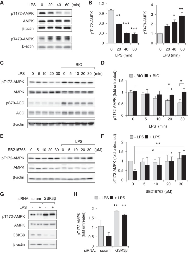
Fig. 3.
GSK3β contributes to dephosphorylation of pT172-AMPK in LPS-stimulated macrophages. A and B: peritoneal macrophages were treated with LPS (300 ng/ml) for the indicated times (0 to 60 min). Representative Western blots and quantitative data show fold changes of pT172-AMPK, pT479-AMPK, total AMPK, and β-actin. Means ± SD (n = 3), *P < 0.05, **P < 0.01, ***P < 0.001, compared with untreated cells. C and D: RAW 264.7 macrophages were pretreated with BIO (0 or 5 μM) for 60 min followed by incubation with LPS (300 ng/ml) for the indicated times. In E and F, cells were incubated with SB216763 (0, 5, 10, 20, or 30 μM) for 60 min and then with or without LPS (300 ng/ml) for an additional 60 min (results were obtained from 2 independent experiments). D and F: means ± SD (n = 3), *P < 0.05, **P < 0.01. G and H: representative Western blots and quantitative data show the amounts of pT172-AMPK in LPS (0 or 300 ng/ml)-treated [scrambled siRNA (scram)] or GSK3β-depleted RAW 264.7 cells. Means ± SD (n = 3), **P < 0.01, compared with untreated or treated with LPS.

