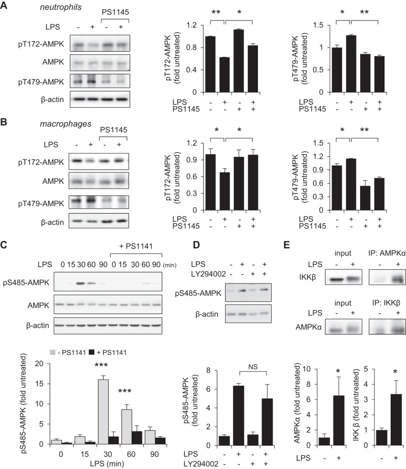
Fig. 5.
IKK and GSK3β participate in inhibiting AMPK phosphorylation in LPS-stimulated neutrophils and macrophages. A and B: bone marrow neutrophils (A) or RAW 264.7 macrophages (B) were treated with the IKK inhibitor PS1145 (0 or 10 μM) for 60 min followed by exposure to LPS (0 or 300 ng/ml) for 60 min. Western blots of pT172-AMPK, pT479-AMPK, AMPK, and β-actin and quantitative data are shown. Means ± SD (n = 3), *P < 0.05, **P < 0.01. C: Western blot analysis show the time-dependent increase in Ser485-AMPK phosphorylation in LPS (300 ng/ml)-treated RAW 264.7 cells. IKK inhibitor PS1141 (10 μM) was applied 60 min prior to LPS exposure. Means ± SD (n = 3), ***P < 0.001. D: RAW 264.7 cells were subsequently cultured with LY294002 (0 or 10 μM) for 60 min followed by inclusion of LPS (0 or 300 ng/ml) for an additional 60 min. Representative Western blots and quantitative are shown (means ± SD, n = 3). E: RAW 264.7 cells were cultured with or without LPS (300 ng/ml) for 30 min followed by pull-down assay with anti-AMPKα or anti-IKKβ antibody. Western blots show the amounts of AMPKα and IKKβ in cell lysates and after immunoprecipitation. Means ± SD (n = 3), *P < 0.05, **P < 0.01.

