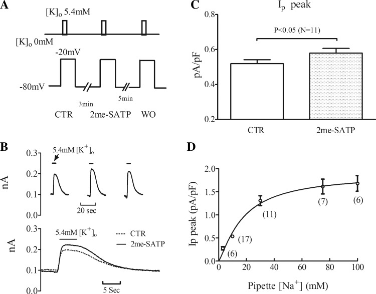
Fig. 1.
Peak Na+-K+-ATPase current (Ip) increases after 2-methylthio-ATP (2-meSATP) application in P2X4 receptor-overexpressing transgenic (P2X4R TG) cardiac myocytes. A, top: protocol of the rapid solution changes with 5.4 mM extracellular K+ concentration ([K+]o) to elicit K+-activated Ip. Bottom, membrane voltage. Ip was recorded at −20 mV, and 2-meSATP was applied at −80 mV. CTR, control; WO, washout. B, top: individual traces in a typical myocyte showing the increase of [K+]o-activated Ip after 2-meSATP application. Bottom, superimposed traces of Ip under CTR conditions and with 2-meSATP. C: peak Ip, normalized by cell capacitance, is presented as means ± SE. Average CTR peak Ip differed from that with 2-meSATP (n = 11 myocytes from 9 TG mice, P < 0.05). D: relationship between peak Ip and pipette Na+ concentration ([Na+]). Peak Ip was elicited by switching [K+]o from 0 to 5.4 mM at −20 mV; pipette [Na+] was varied as shown. Normalized peak Ip is presented as means ± SE. Numbers of cells are indicated in the parentheses (from 18 TG mice).

