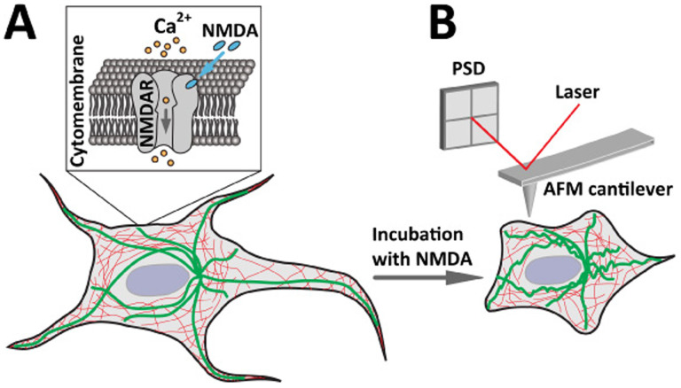
Figure 1. Schematic diagrams showing the NMDA induced structural changes of live cell that can be detected by AFM.
(A) The process of the cell response under NMDA treatment. NMDA specifically binds to the NMDA receptors (NMDAR) in the cytomembrane that opens the ligand gated ion channel to facilitate Ca2+ influx into the cell. (B) AFM measurement of live cell response under NMDA treatment. NMDA treatment and Ca2+ influx induce reorganization of cytoskeleton (thick and green lines indicate microtubules, thin and red lines indicate actin filaments) and the resulting structural changes of the cell can be detected by AFM in high resolution and real time.

