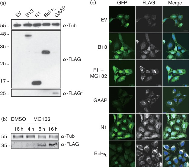
Fig. 1.
Expression of VACV anti-apoptotic proteins by lentivirus-transduced U2-OS cells. (a) Lysates of lentivirus-transduced U2-OS cells were analysed by SDS-PAGE and immunoblotting with indicated antibodies. A long exposure is indicated with an asterisk. (b) For F1 detection, the cells were treated with MG132 for the indicated time prior to immunoblotting. Tub, α-tubulin. Molecular size markers are indicated on the left (kDa). (c) Immunofluorescence. Cells were fixed and stained for immunofluorescence analysis using rabbit anti-FLAG antibody. The expression of GFP, FLAG-tagged proteins and merged images with DNA stained with DAPI are shown. Bar, 20 µm.

