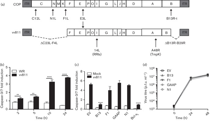
Fig. 4.
vv811 infection of U2-OS cell lines expressing VACV anti-apoptotic proteins. (a) Schematic of the genome structure of VACV strains COP and vv811, with the position of the anti-apoptotic proteins indicated. ITR, inverted terminal repeat; B13R-t, truncated B13R gene; RRls, ribonucleotide reductase large subunit; TmpK, thymidylate kinase. Note the C12L gene of VACV strain COP is equivalent to the B22R gene of VACV strain WR. (b) U2-OS parental cells were infected at 2.5 p.f.u. per cell with WR or vv811 and caspase-3/7 activity was assessed when indicated using Caspase-Glo. Data were normalized to mock-infected cells to obtain a fold induction. (c) U2-OS cell lines were infected with vv811 at 2.5 p.f.u. per cell for 24 h and apoptosis was assessed quantitatively using Caspase-Glo. Data are shown as the mean±sd and are representative of at least three experiments performed in triplicate. Statistical differences were determined using an unpaired Student’s t-test (*P<0.05, **P<0.01, ****P<0.0001). (d) Cells were infected with vv811 at 0.1 p.f.u. per cell, harvested at the indicated time and the intracellular virus titre was determined by plaque assay on BSC-1 cells.

