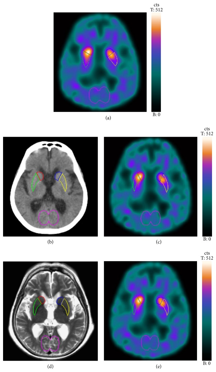
Figure 1.
Regions of interest (ROIs) were manually drawn directly on the summed SPECT image (a). In CT-guided method, ROIs were manually delineated directly on one CT slice with the best recognizable striatum. ROIs were then transferred to the hardware-based coregistered summed SPECT image (b, c). In MRI-guided method, ROIs were manually delineated directly on one MRI slice with the best recognizable striatum. ROIs were then transferred to the software-based coregistered summed SPECT image (d, e).

