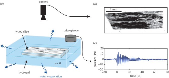
Figure 2.
Experiment set-up: (a) the water contained in the wood inclusion in hydrogel underwent negative pressure as water evaporated from the hydrogel. The camera installed on an optical microscope with magnification between 1.25× and 20× recorded images (b) of bubble development (black conduits on image) in the water columns (grey on image). (c) Simultaneously, the microphone monitored the ultrasonic signals. (Online version in colour.)

