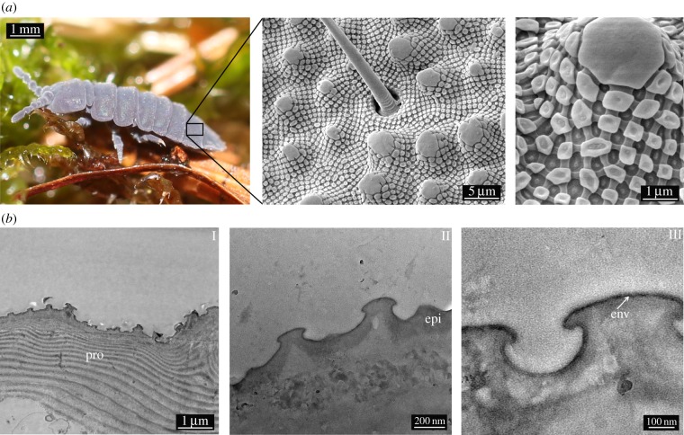
Figure 1.
(a) SEM studies of the cuticular morphology of T. bielanensis. SEM images showing papillose microstructures (secondary granules), covered by a rhombic comb-like mesh exhibiting nanoscopic tubercles (primary granules). (b) TEM sections of the cuticle, revealing the layered structure to consist of (I) the lamellar procuticle (pro), covered by (II) the epicuticle (epi) and (III) a thin envelope (env) as the topmost layer. (Online version in colour.)

