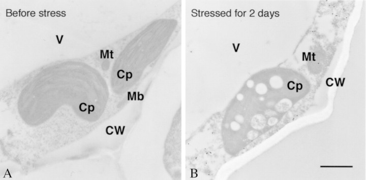
Fig. 5. Immunoelectron microscopy of wild watermelon leaves before and after drought stress for 2 d with the anti‐DRIP‐1 antibody. Samples were prepared for immunoelectron microscopy as described by Seo et al. (2000). Sections were incubated with anti‐DRIP‐1 antibody raised in rabbits to the synthetic peptide (Tyr‐248 to Glu‐263 of DRIP‐1) and then reacted with anti‐rabbit IgG conjugated with 20 nm diameter particles (Biocell Research Laboratories, Cardiff, UK). After immunolabelling, the sample was stained with uranyl acetate. Bar = 1 µm. CW, Cell wall; Cp, chloroplast; Mb, microbody; Mt, mitochondrion; V, vacuole.

An official website of the United States government
Here's how you know
Official websites use .gov
A
.gov website belongs to an official
government organization in the United States.
Secure .gov websites use HTTPS
A lock (
) or https:// means you've safely
connected to the .gov website. Share sensitive
information only on official, secure websites.
