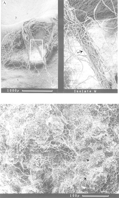
Fig. 1. A, Scanning electron micrographs of bean cotyledon surface colonized by mycelium of Hebeloma syrjense (x). Rhizomorph‐like structures (boxed, and enlarged in B and arrowed) can be seen extending from the original inoculum plug (y) to the cotyledon surface densely covered by mycelium. C, SEM of interior of bean cotyledon invaded by mycelium of H. syrjense. Individual cells (arrowed bulbous structures) are surrounded and obscured by a dense net of hyphae.
