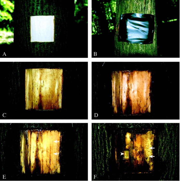
Fig. 1. Macroscopic view of a wound surface in lime. A, Fresh wound; B, wound covered with black plastic wrap; C, discolouration on some parts of the wound surface after 1 week; D, intense discolouration in the centre and on the left‐hand side of the wound after 2 weeks; E, 3 weeks after wounding a bright callus tissue develops (arrows); F, after 9 weeks nearly half the wound is covered with bright surface callus tissue (arrows); arrowhead indicates place of sampling for LM and TEM.
