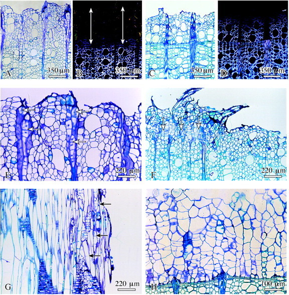
Fig. 2. Early stage of surface callus development in lime. A, C, E–H, Bright‐field microscopy; B and D, polarized light. A and B, Layer of xylem cells without birefringence as a marker for missing secondary wall (arrows); C and D, nearly all cells on the wound surface show birefringence under polarized light showing that these cells have already formed a secondary wall; E, proliferation and cell division of the ray parenchyma cells (arrows); F, young callus tissue (arrows) on parts of the wound surface; G, formation of transverse walls in fibres close to the wound surface (arrows); H, complete layer of surface callus cells directly attached to previous year’s latewood. A–F and H, Transverse sections; G, radial.
