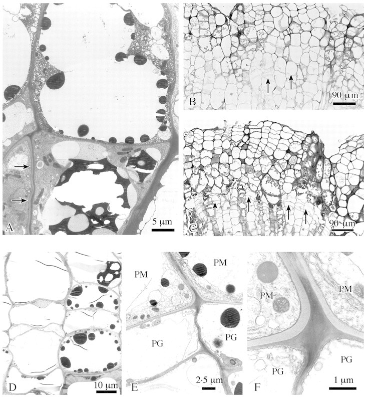
Fig. 4. Formation of wound periderm in lime. A, In outer callus cells suberin‐like layers are attached to the cell wall (arrows) and dark staining substances are deposited in vacuoles. B and C, A phellogen develops inside the outer callus tissue, some cells insert tangential walls (arrows) (B); later a complete tangential layer of flattened phellogen cells is formed (arrows) (C). D, Detail of phellogen formation: isodiametric callus cells change into flattened cells by insertion of tangential walls. E, Young phellem cells (PM) with suberized additional wall layer and phellogen cells (PG). F, Detail of E. Phellogen (PG) and phellem (PM) cells, the latter with suberin‐like layer being formed. Transverse sections; B and C, LM; A, D–F, TEM.
