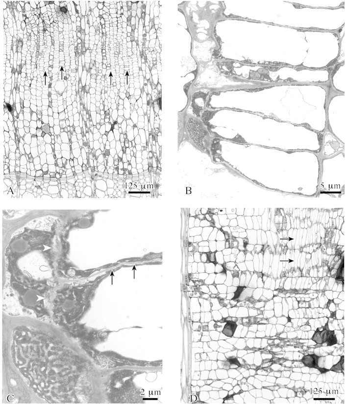
Fig. 5. Wound cambium formation within surface callus tissue in lime. A, A new wound cambium develops (arrows) close to previous year’s xylem. B, Radially flattened wound cambium cells develop from isodiametric callus cells by insertion of tangential and radial walls. C, Detail of B showing formation of tangential (arrow) and radial (arrowheads) walls. D, Cambial cells are axially elongated (arrows) as compared with callus cells. A–C, transverse sections; D, radial section. A and D, LM; B and C, TEM.
